Baker's Cyst Aspiration
KneeEquipment
Medications
Standard pre-procedure workup: consent, indications, contraindications, allergies, and baseline pain level.
Position patient prone with the knee in slight flexion (pillow under ankle). The prone position brings the popliteal fossa to the surface and provides the easiest access to the cyst.
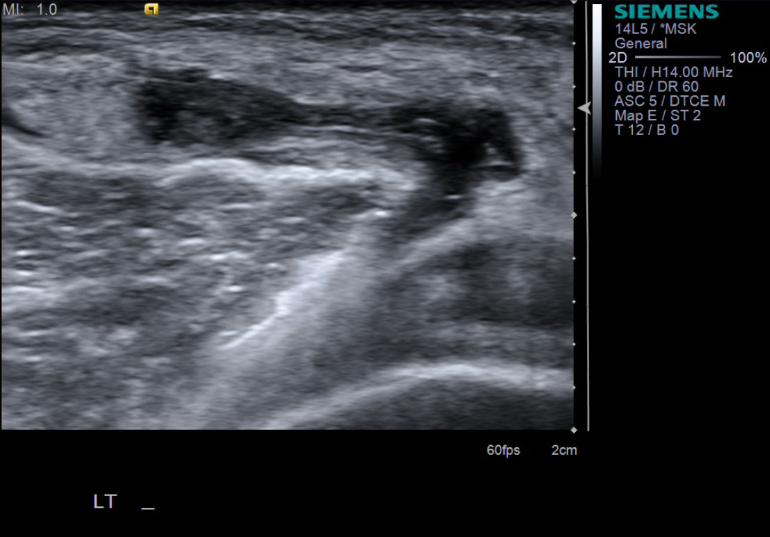
Scan the popliteal fossa with the linear probe in the transverse plane. Identify the Baker's cyst (popliteal cyst) arising between the medial head of the gastrocnemius and the semimembranosus tendon. Measure the cyst in two planes and assess for internal debris, septations, or rupture. Identify the popliteal artery and vein (medial and central in the fossa) using color Doppler.


Mark the needle entry site. Plan a medial approach to avoid the popliteal neurovascular bundle. The needle typically enters from the medial side of the cyst.
Prep with betadine or chlorhexidine × 3.
Place sterile drape and sterile probe cover.
Under ultrasound guidance, inject 1% lidocaine superficially using the 25g needle. Anesthetize down to the cyst wall.
Exchange for the 18g needle attached to the 20 cc aspiration syringe. Advance under real-time ultrasound guidance into the cyst body. Confirm needle tip position within the cyst before aspirating.
Aspirate the cyst contents. The fluid is typically viscous and yellow-brown. Advance the needle further into the cyst as it collapses to aspirate residual fluid. Aspirate as much fluid as possible.
Once aspiration is complete, disconnect the 20 cc syringe while leaving the 18g needle in place. Exchange for the 3 cc steroid/ropivacaine syringe and inject into the residual cyst cavity.

Scan post-procedure to document cyst decompression and confirm no hematoma formation.
Remove needle, apply pressure to the entry site, clean with alcohol, and place bandage.
Advise patient that the cyst may recur if the underlying intra-articular pathology (e.g., osteoarthritis, meniscal tear) is not addressed. Reassess pain level and provide a pain log.
References
- Smith MK, Lesniak B, Baraga MG, Kaplan L, Jose J. Treatment of popliteal (Baker) cysts with ultrasound-guided aspiration, fenestration, and injection: long-term follow-up. Sports Health. 2015;7(5):409–414.
- Di Sante L, Paoloni M, Ioppolo F, Dimaggio M, Di Renzo S, Santilli V. Ultrasound-guided aspiration and corticosteroid injection of Baker's cysts in knee osteoarthritis: a prospective observational study. Eur J Phys Rehabil Med. 2012;48(4):561–567.
- Köroğlu M, Callıoğlu M, Eriş HN, et al. Ultrasound guided percutaneous treatment and follow-up of Baker's cyst in knee osteoarthritis. Eur J Radiol. 2012;81(11):3466–3471.