Radiocapitellar Joint Injection
ElbowEquipment
Medications
Standard pre-procedure workup: consent, indications, contraindications, allergies, and baseline pain level.
Position patient seated or supine with the elbow flexed to approximately 90° and the forearm pronated, resting on the patient's lap or a pillow.
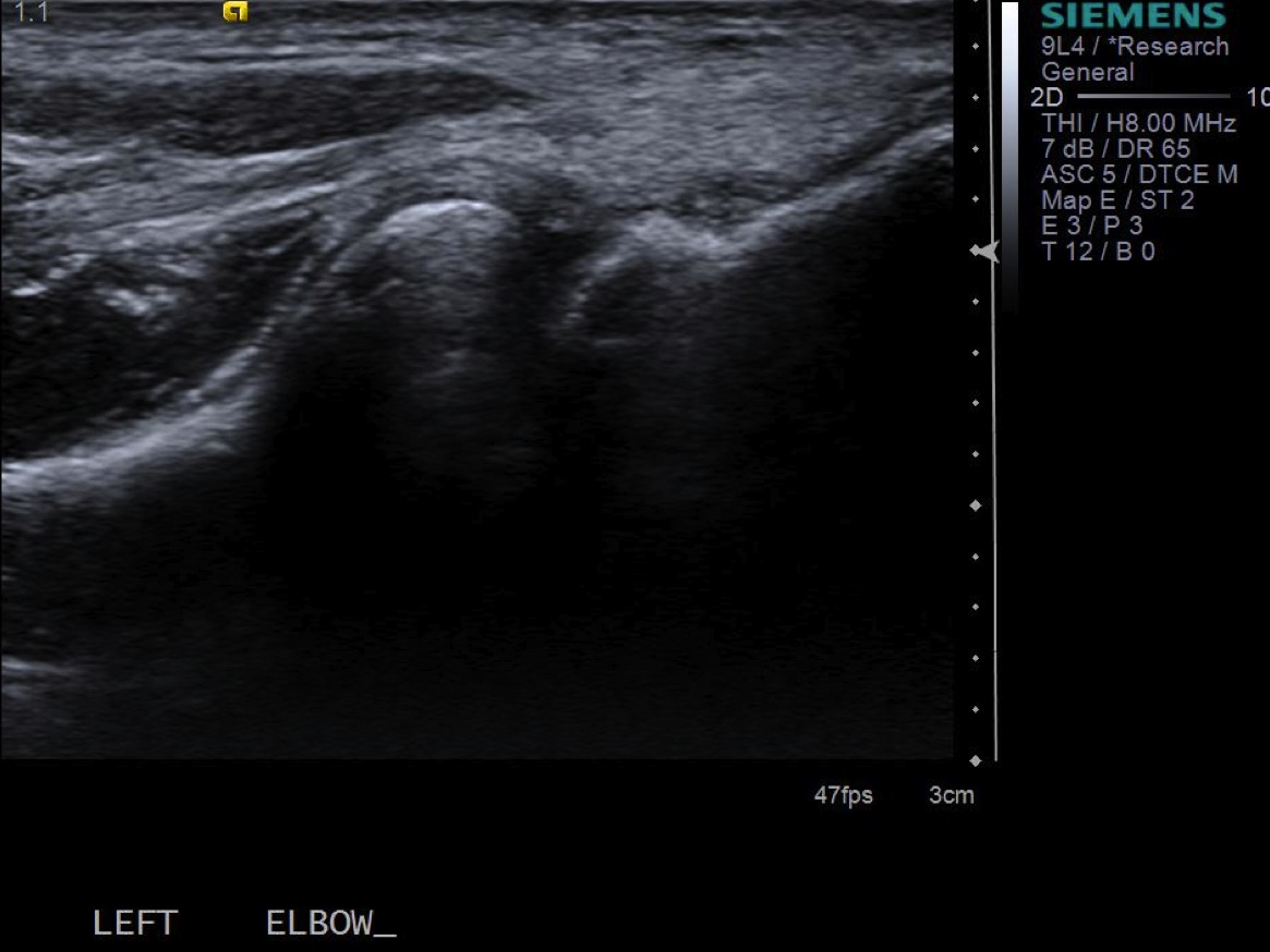
Place the linear probe over the lateral elbow in the long axis of the radius (coronal plane). Identify the radiocapitellar joint — the radial head articulating with the capitellum of the humerus.

Assess for joint effusion or synovitis in the posterior radiocapitellar recess.
Mark the needle entry site and ideal probe position on the skin.
Prep with betadine or chlorhexidine × 3.
Place sterile drape and sterile probe cover.
Under ultrasound guidance, create a skin wheal with 1% lidocaine using the 25g needle. Advance into the subcutaneous tissues.

Advance needle under ultrasound guidance into the radiocapitellar joint space. The needle tip should be positioned between the radial head and the capitellum.


Test-inject with 1% lidocaine. If there is no resistance and you see joint distention, disconnect the syringe and exchange for the steroid/ropivacaine mixture. Inject slowly.
Remove needle, clean skin with alcohol, and place bandage.
Reassess pain level and provide patient with a pain log.
References
- Lin JS, Gimarc DC, Adler RS, et al. Ultrasound-guided musculoskeletal injections. Semin Musculoskelet Radiol. 2021;25(6):769–784.
- Lapegue F, André A, Lafourcade F, et al. Ultrasound of lateral epicondylitis. Semin Musculoskelet Radiol. 2024;28:683–693.